At Southern Imaging Services (SIS), we specialize in advanced neurological imaging with cutting-edge technology. Whether you need a brain MRI, spine MRI, or cranial nerve imaging, you can rely on us for accurate diagnosis and quick results. Our board-certified radiologists will submit MRI imaging results to the treating physician within 24 to 48 hours.
We offer non-contrast MRI studies with cutting-edge technology and a patient-first approach. Whether you visit us in Charlotte, High Point, Raleigh, Wilmington, or Rock Hill, you can count on us to provide excellent care in a welcoming environment. Our team speaks fluent English and Spanish.
Book your MRI today.
Neurological Imaging at SIS
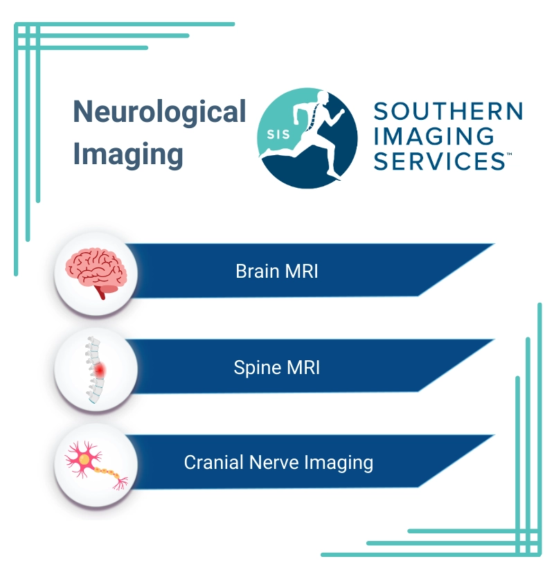
At Southern Imaging Services (SIS), we offer high-quality, non-contrast MRI imaging of the brain, spinal cord, and nerves.
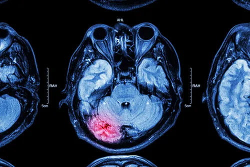
Brain MRI
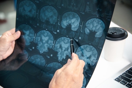
A brain MRI is a non-invasive neurological imaging that provides detailed pictures of the brain’s structure. At Southern Imaging Services, we use non-contrast brain MRI scans to evaluate various neurological conditions.
A brain MRI can detect signs of stroke, revealing areas of damaged brain tissue. It’s also excellent for identifying brain tumors, assessing traumatic brain injuries, and monitoring the progression of multiple sclerosis. MRI brain imaging helps diagnose and track neurodegenerative diseases such as Alzheimer’s and Parkinson’s, showing changes in brain structure over time
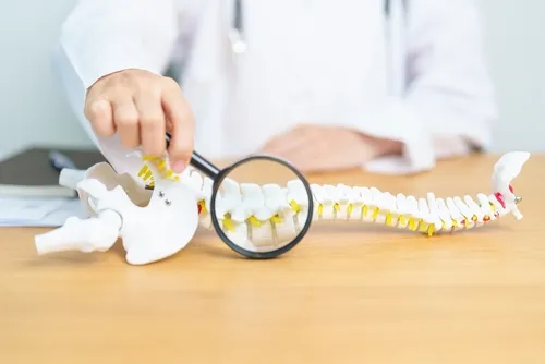
Spine MRI
Spine MRI imaging is a valuable diagnostic test for back pain and other spinal problems. A spine MRI offers a clear view of the spinal cord, vertebrae, and surrounding tissues. It’s especially helpful for detecting spinal cord compression, which can cause pain and neurological symptoms.
MRI scans of the spine can also identify disc herniations, where the soft inner material of a spinal disc protrudes and potentially irritates nearby nerves. They are also useful for diagnosing degenerative disc disease, a condition where the spinal discs wear down over time, leading to pain and reduced mobility.
Cranial Nerve Imaging
Cranial Nerve Imaging utilizes high-resolution MRI techniques to visualize the twelve pairs of cranial nerves that control various functions in the head and neck. Detailed images of these delicate structures guide treatment decisions for various neurological conditions.
Cranial Nerve Imaging is used to diagnose disorders such as trigeminal neuralgia, a condition causing severe facial pain. It’s also crucial for detecting vestibular schwannomas, also known as acoustic neuromas. These are benign tumors that can affect hearing and balance.
At SIS, your well-being is our priority. With convenient locations and a dedicated team, we’re committed to providing you with the information and care you need to support your health journey. Whether you require neurological imaging for diagnosis, treatment monitoring, or preventive care, SIS is here to help.
How MRI Neuroimaging Works
Magnetic Resonance Imaging (MRI) is a safe and non-invasive way to create detailed pictures of the inside of your body. It uses a powerful magnet, radio waves, and a computer to take pictures of your brain, spine, or cranial nerves. Unlike X-rays or CT scans, MRI does not use radiation, making it a safer option for many people. The images produced by an MRI are incredibly detailed, allowing doctors to see structures in your body with great clarity and diagnose or monitor medical conditions effectively.
During an MRI scan, the strong magnet aligns the hydrogen atoms in your body, which are naturally present in water and fat. Then, the MRI machine sends radio waves into your body, causing these atoms to produce signals. The machine collects these signals and a computer processes them to create detailed images of your internal organs and tissues. Non-contrast MRI scans mean no dye or injections are needed, making the process even more comfortable and simple for patients.
What to Expect From an MRI Scan
At Southern Imaging Services, we strive to make your MRI experience as smooth and comfortable as possible, providing the answers you need for your health journey.
Before Your Scan
We recommend arriving early and wearing comfortable clothes without metallic buttons or zippers. However, we will offer scrubs to change into if needed.
Before your scan, you will be asked to remove metal objects such as glasses, jewelry, dentures or dental bridges, and body piercings because the strong MRI magnet can affect certain metals.
It’s also important to let the staff know if you have any metal implants or medical devices in your body. They will assess if it’s safe for you to undergo the MRI.
What Happens During the MRI Scan
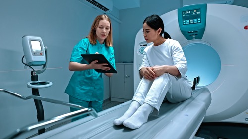
Once you’re ready, you will lie on a padded table that slides into the MRI machine, which looks like a large tube. The area of your body being scanned, whether it’s your brain, spine, or cranial nerves, will be positioned in the center of the machine.
The scan is painless, but the machine makes loud tapping and thumping noises. To make you more comfortable, you may be given earplugs or headphones to block out the noise. It’s important to stay still during the scan so that the images come out clear.
How Long the MRI Takes
Each MRI should take less than 30 minutes. When physicians order multiple MRIs, patients may take breaks between procedures. Our staff will communicate with you throughout the test, and you can let us know if you need assistance.
After Your MRI Scan
When the scan is complete, the table will slide out of the machine, and you can go about your day as usual. There are no side effects or recovery time since no contrast dye is used. The images from your scan will be sent to a radiologist, a doctor who specializes in reading and interpreting medical images. Your healthcare provider will then review the results with you and discuss any next steps if needed.
Neurological Imaging FAQs (Frequently Asked Questions)
How Soon Will I Get My MRI Results?
One of our experienced Board Certified Radiologists will read your MRI in real-time and send it to your referring physician within 24-48 hours. Your physician will then review and discuss your MRI results and treatment options with you.
How Much Does a Brain MRI, Spine MRI, or Nerve MRI Cost?
We are committed to offering high-quality neurological imaging at the lowest possible cost.
Unlike other providers, we will charge a self-pay price at the time of service. You will not be surprised by hidden charges, such as radiologist reading fees or facilities fees. As a self-pay patient, you’ll pay for your MRI at the time of service with no surprise bills in your mailbox later.
The Self-Pay Price Includes:
- Your MRI scan
- A copy of your exam on a CD-ROM
- Professional interpretation and a written report from a board-certified radiologist sent to you and/or your physician within 24-48 hours of your exam.
For pricing please call 704-321-4798.
Why Choose Southern Imaging Services?
- Highest Quality Imaging: SIS provides the highest quality MRI imaging with state-of-the-art technology for accurate results.
- Experienced Staff: Our radiologists are all board-certified with sub-specializations in Musculoskeletal (MSK) or Neuroradiology.
- Quick Results: MRI results will be sent to your referring physician within 24-48 hours.
- English and Spanish: ¿hablas español? Our team is fluent in both English and Spanish.
Contact SIS Today to Schedule Your Neuroimaging
At Southern Imaging Services, we’re committed to providing safe, accurate, and convenient MRI imaging to help you better understand and manage your health. Whether you need a brain MRI, spine MRI, or cranial nerve imaging, you can rely on our experienced team at SIS.
With multiple locations in North Carolina and bilingual staff fluent in English and Spanish, we make it easy to access the care you need. Don’t wait, contact us today to schedule your appointment and take the next step toward a healthier you.