Magnetic Resonance Imaging (MRI) is an essential tool in neurology, giving us a look into the brain and spinal cord like never before. This technology uses powerful magnetic fields and radio waves to create detailed images of soft tissues, making it a vital diagnostic tool for many neurological conditions. From multiple sclerosis, brain tumors, and strokes to treatment plans and surgical interventions, MRI has changed the way we approach neurological care. Beyond the role of MRI in diagnosing neurological disorders in clinical use, MRI is also crucial in research, allowing scientists to understand brain activity, disease progression, and treatment effects.
In this blog, we explore the role of MRI in diagnosing neurological disorders and the importance of visiting our Charlotte imaging center for state-of-the-art imaging services.
MRIs and Their Significance
Magnetic Resonance Imaging (MRI) is a cornerstone of modern medical imaging, offering unparalleled detail and safety for diagnosing and managing various conditions.
How MRI Works
Magnetic Resonance Imaging (MRI) is a key technology in the medical field. It uses strong magnetic fields and radio waves to create detailed images of the body’s organs and tissues. MRI is particularly valuable for examining the brain and spinal cord. This imaging technique provides high-resolution images, allowing healthcare professionals to visualize complex structures and detect abnormalities.
Safety Advantages of MRI
One significant advantage of MRI is its safety. Unlike computed tomography (CT) scans, MRI does not use ionizing radiation. This makes it a safer option for patients, especially for those requiring multiple scans over time. MRI technology minimizes exposure to harmful radiation, making it suitable for various patient populations, including children and pregnant women.
Role of MRI in Diagnosing Neurological Disorders
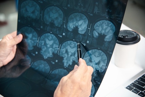
The ability to produce detailed images enhances the role of MRI in diagnosing neurological disorders. By providing clear views of brain structures, MRI allows for the identification of conditions such as brain tumors, multiple sclerosis, and traumatic brain injuries. This technology is essential for accurate diagnosis and effective treatment planning.
Types of MRI and Their Applications
Magnetic Resonance Imaging (MRI) is a versatile tool in the diagnosis of neurological disorders. Different types of MRIs provide unique insights into brain structure and function. Here are the main types of MRI and their specific applications:
Conventional MRI
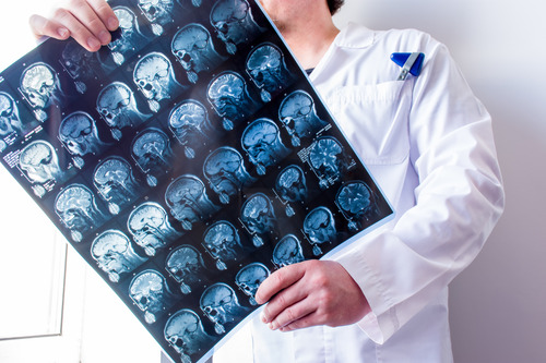
Conventional MRI is primarily used for structural imaging. It provides detailed images of the brain and spinal cord. This type of MRI is crucial for identifying conditions such as brain tumors and strokes. The high-resolution images allow healthcare professionals to see the size, shape, and location of abnormalities. This information is vital for accurate diagnosis and treatment planning.
Functional MRI (fMRI)
Functional MRI (fMRI) measures brain activity by detecting changes in blood flow. It is particularly useful in mapping brain activity during specific tasks. This capability makes fMRI valuable in pre-surgical planning. Surgeons can identify critical areas of the brain that control essential functions, helping to avoid damage during procedures. fMRI also aids in understanding neurological conditions by revealing how different regions of the brain communicate.
Diffusion Tensor Imaging (DTI)
Diffusion Tensor Imaging (DTI) is a specialized MRI technique that visualizes white matter tracts in the brain. It measures the movement of water molecules in brain tissue, providing insights into the integrity of white matter. DTI is beneficial for diagnosing conditions such as multiple sclerosis and traumatic brain injuries. It helps in assessing the impact of these disorders on brain connectivity.
Magnetic Resonance Spectroscopy (MRS)
Magnetic Resonance Spectroscopy (MRS) offers a chemical analysis of brain tissue. Unlike conventional MRI, which focuses on structure, MRS evaluates the chemical composition of the brain. This technique can detect metabolic changes associated with various neurological disorders. For example, MRS can help identify abnormalities in brain metabolism linked to tumors or neurodegenerative diseases.
Diagnosing Neurological Disorders
Magnetic Resonance Imaging (MRI) plays a crucial role in diagnosing various neurological disorders. Its ability to produce high-resolution images of the brain and spinal cord allows healthcare professionals to identify conditions early and accurately.
Early Detection and Monitoring of Brain Tumors
MRI is essential for the early detection of brain tumors. It provides detailed images that help doctors assess the size and location of tumors. This information is vital for determining the best treatment options. Regular MRI scans also allow healthcare providers to monitor tumor progression or response to therapy effectively.
Identifying Ischemic and Hemorrhagic Strokes
MRI is a key tool in identifying strokes. It can distinguish between ischemic strokes, caused by blocked blood flow, and hemorrhagic strokes, caused by bleeding in the brain. MRI scans can reveal the extent of brain damage and help medical teams decide on immediate treatment strategies. Early detection through MRI significantly improves patient outcomes.
Role of MRI in Diagnosing and Tracking Multiple Sclerosis
Multiple sclerosis (MS) is a complex neurological disorder that affects the central nervous system. MRI is critical in diagnosing MS by identifying lesions in the brain and spinal cord. It also plays a vital role in tracking the progression of the disease. Regular MRI scans can help assess the effectiveness of treatment and monitor any new lesions.
Assessing Traumatic Brain Injuries with Detailed Images
Traumatic brain injuries (TBIs) can have severe consequences. MRI provides detailed images that help diagnose the extent of brain injuries. It can detect subtle changes in brain structure that may not be visible in other imaging modalities. This information is essential for developing an appropriate treatment plan and predicting long-term outcomes for patients.
Monitoring Treatment Progress
The role of MRI in diagnosing neurological disorders allows for proper monitoring of treatment progress. Repeated MRI scans provide valuable information about how well a treatment is working. This process allows healthcare professionals to make informed decisions about patient care.
Importance of Repeated MRI Scans in Tracking Treatment Effectiveness
Regular MRI exams help doctors assess the effectiveness of treatments. For example, in patients with brain tumors, MRI scans can show changes in tumor size. If the tumor shrinks, it indicates that the treatment is working. Conversely, if the tumor remains the same or grows, doctors may need to adjust the treatment plan.
In conditions like multiple sclerosis, MRI scans reveal new lesions or changes in existing ones. Tracking these changes helps doctors evaluate how well a patient responds to medication. Early detection of new lesions can prompt timely adjustments to treatment, which can improve patient outcomes.
Clinical and Research Applications
Magnetic Resonance Imaging (MRI) has revolutionized the field of neurology in both clinical diagnosis and groundbreaking research.
MRI in Clinical Settings for Diagnosis and Treatment Planning
Magnetic Resonance Imaging (MRI) plays a vital role in clinical settings. Healthcare professionals use MRI to diagnose various neurological disorders. MRI provides detailed images of the brain and spinal cord, allowing for accurate assessments. This imaging technology helps identify conditions such as brain tumors, strokes, and multiple sclerosis.
For instance, when a patient presents with symptoms of a stroke, an MRI can quickly reveal whether it’s ischemic or hemorrhagic. This distinction is crucial for determining the appropriate treatment. Additionally, MRI assists in treatment planning. Surgeons rely on MRI images to visualize the affected areas before performing operations. This preparation enhances surgical precision and improves patient outcomes.
Contributions to Research in Understanding Neurological Conditions
The role of MRI in diagnosing neurological disorders is not only essential in clinical practice but also in research. Researchers use MRI to study the brain’s structure and function. This technology allows scientists to explore how neurological disorders develop and progress. For example, studies utilizing functional MRI (fMRI) have shed light on brain activity patterns in patients with epilepsy.
Moreover, the role of MRI in diagnosing neurological disorders contributes to understanding the effects of various treatments on the brain. Researchers can monitor changes over time, providing valuable data on treatment efficacy. This information is crucial for developing new therapies and improving existing ones.
Visit Our Charlotte Imaging Center For Your Diagnostic Needs!
If you’re looking for advanced imaging services, trust our dedicated team at Southern Imaging Services to deliver accurate results with care. Using state-of-the-art MRI technology, we provide detailed insights to support your diagnosis and treatment planning. Our compassionate professionals are committed to ensuring your comfort and answering any questions you may have throughout the process.
Contact us at (704) 321-4798 to schedule an appointment with us today!