Understanding Brain Magnetic Resonance Imaging
A Brain MRI, or magnetic resonance imaging for the brain, is a specialized imaging test that provides detailed pictures of brain structures. This non-invasive procedure uses powerful magnets and radio waves to create high-quality images of the brain and spinal cord. Unlike other imaging techniques, such as CT scans, an MRI for brain scans does not utilize ionizing radiation, making it a safer option for many patients.
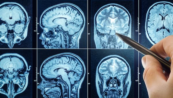
During the exam, the MRI machine generates a magnetic field that interacts with the water molecules in your body. This interaction allows the MRI scanner to produce clear images of brain tissue, blood vessels, and surrounding structures. The resulting images help doctors diagnose various neurological conditions, assess brain injuries, and evaluate abnormalities such as tumors or lesions.
Brain MRIs are particularly effective for visualizing soft tissues. They can reveal critical details about brain tissue, blood flow, and even changes associated with neurological disorders like multiple sclerosis or Alzheimer’s disease. This makes the Brain MRI an essential tool for healthcare providers in diagnosing and monitoring conditions affecting the brain.
Understanding the purpose and process of a Brain MRI can help alleviate any concerns you may have. If you are scheduled for a Brain MRI, rest assured that you will receive precise and accurate imaging that will aid your healthcare provider in making informed decisions regarding your treatment and care.
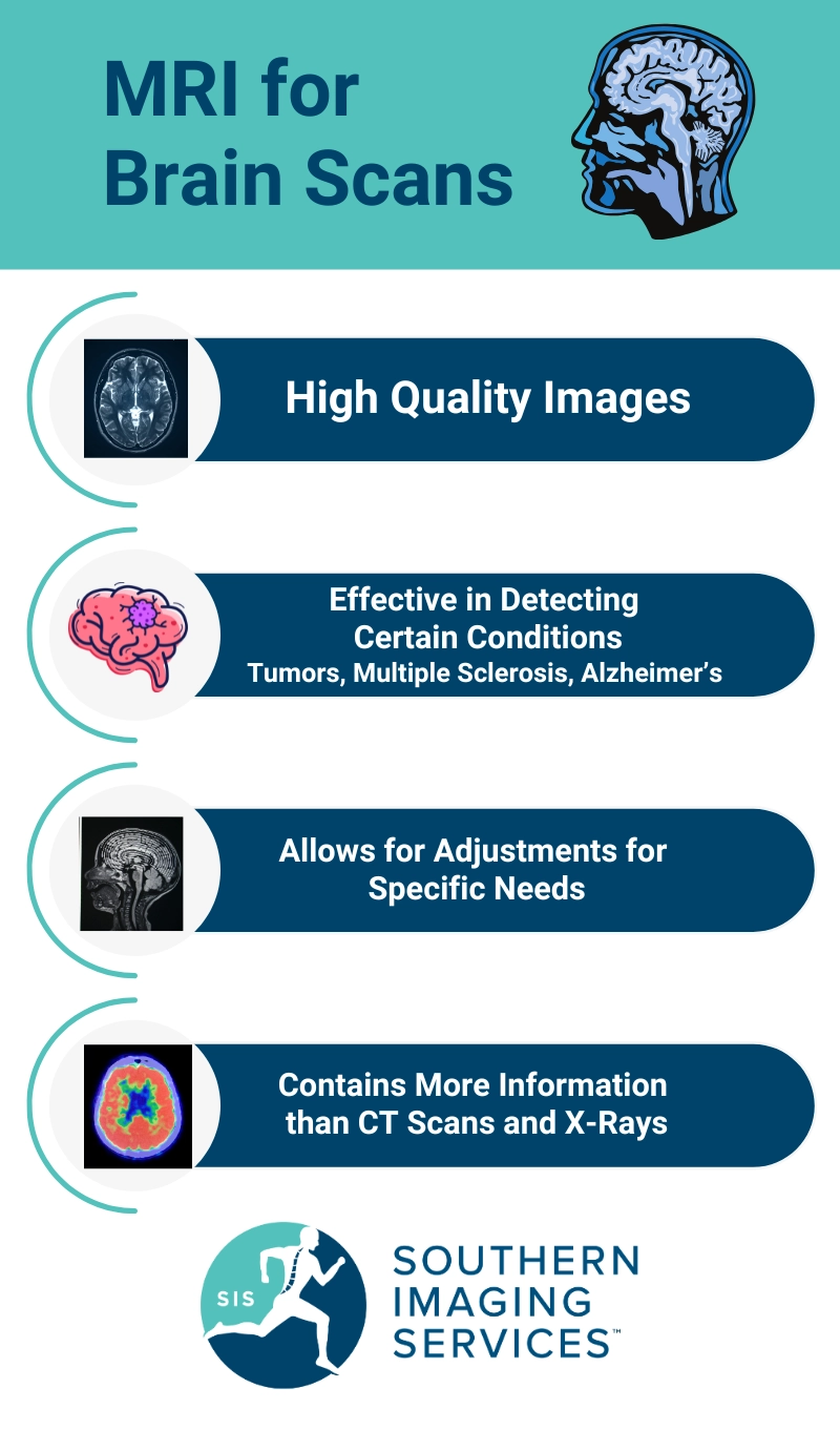
Why Choose MRI for Brain Imaging?
Magnetic Resonance Imaging (MRI) is a powerful tool for examining the brain. It provides detailed images that help in diagnosing various neurological conditions. Choosing an MRI for brain imaging offers several advantages.
First, MRI uses a strong magnetic field and radio waves to create high-quality images of brain structures. Unlike X-rays or CT scans, MRI does not involve ionizing radiation, making it a safer option for patients. The detailed pictures produced by the MRI can reveal abnormalities in brain tissue, blood vessels, and surrounding tissues, which are crucial for accurate diagnoses.
Second, brain MRI is particularly effective for detecting conditions such as brain tumors, multiple sclerosis, and brain damage. It allows healthcare providers to visualize the brain and spinal cord in great detail, aiding in the assessment of neurological disorders. This imaging test is also useful for monitoring the progression of diseases like Alzheimer’s disease and evaluating the effects of brain surgery.
Additionally, an MRI for brain scans can be tailored to specific needs. For instance, functional MRI (fMRI) can assess brain activity by measuring blood flow, providing insights into how different brain regions work together. This is especially beneficial for planning surgical interventions or rehabilitation strategies.
Since MRI produces highly detailed images, allowing for the visualization of fine structures within the brain, it can be seen as a sensitive imaging test that can detect things X-rays and CT (computed tomography) scans can’t. A head MRI is one of the most direct ways to detect brain injuries or diseases.
In summary, choosing an MRI for brain imaging offers a comprehensive view of brain health. Its ability to produce clear images without radiation exposure makes it an excellent choice for patients seeking precise diagnoses and effective treatment plans.
Preparing for Your MRI Scan
Preparing for a brain MRI is crucial for obtaining accurate results. Here’s how to get ready for your MRI exam.
How to Prepare for a Brain MRI Exam
Before your appointment, inform your healthcare provider about any medical conditions, allergies, or medications you are taking. If you have any implanted medical devices, such as pacemakers or cochlear implants, it’s important to discuss these as they may affect your MRI scan. You may be asked to wear a hospital gown, so avoid clothing with metal zippers or fasteners. Additionally, you should refrain from wearing jewelry or accessories that contain metal.
If your doctor recommends a contrast injection to enhance the MRI images, they will provide specific instructions. You may need to avoid eating or drinking for a few hours before the exam, especially if contrast material is involved.
What to Expect During the Procedure
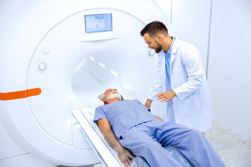
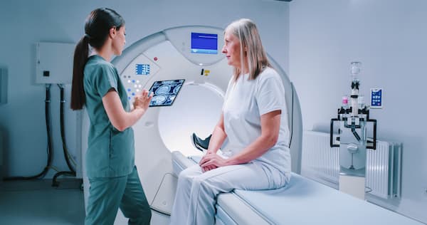
On the day of your MRI scan, arrive at the facility a little early to complete any necessary paperwork. You will be escorted to the exam room and asked to lie down on the MRI unit. The MRI technologist will position you carefully to ensure clear images of your brain structures. Earplugs may be provided to help reduce the noise from the MRI machine, which uses powerful magnets and radio waves to capture detailed pictures of your brain.
During the scan, it is essential to remain still. The procedure typically lasts between 30 to 60 minutes, depending on the complexity of the imaging required. You will be monitored throughout the process, and you can communicate with the technologist if you have any concerns.
Safety Precautions and Considerations
MRI scans are safe and do not involve ionizing radiation. However, you should inform your technologist if you have any claustrophobia, as some MRI machines are enclosed. Open MRI options may be available if you feel anxious in confined spaces.
Always disclose any metal objects in your body, including surgical clips or hearing aids, as these can interfere with the magnetic field. Pregnant individuals should consult their healthcare provider before undergoing an MRI scan.
By following these preparation guidelines, you can help ensure a smooth MRI experience and obtain high-quality images for accurate diagnoses. If you have any questions or concerns, don’t hesitate to reach out to our team at Southern Imaging Services.
Inside the MRI Machine: What Happens During the Scan?
When you arrive for your MRI for brain imaging, you will encounter a sophisticated MRI machine designed to produce detailed images of your brain and spinal cord. The MRI scanner is a large, tube-like structure that uses powerful magnets and radio waves to create high-quality images. You will lie down on a comfortable table that slides into the MRI unit.
During the procedure, it’s important to remain still. Movement can distort MRI images, affecting the quality of the results. The technologist will be in a separate room but will monitor you through a window and maintain communication via intercom. You may also be given earplugs or headphones to help reduce the noise of the MRI machine, which can be quite loud during the scan.
In some cases, contrast material may be used to enhance the images. This contrast dye helps to highlight specific areas of the brain, allowing for clearer visualization of blood vessels and soft tissues. If a contrast injection is necessary, the MRI technologist will explain the process and any potential side effects, such as a temporary metallic taste or allergic reactions.
The entire MRI exam typically lasts between 30 to 60 minutes, depending on the complexity of the images needed. After the scan, you will be able to return to your normal activities right away, as there are no side effects from the MRI itself. Understanding this process can help ease any anxiety you may have about your MRI for brain imaging.
Interpreting Your MRI Results
After your MRI scan, understanding the results is crucial for your health and well-being. The images captured during your brain MRI provide essential insights into various conditions affecting the brain and spinal cord. Here’s a guide to help you navigate the interpretation of your MRI results.
Who Analyzes MRI Images?
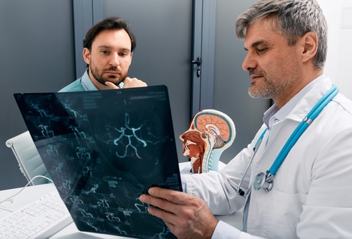
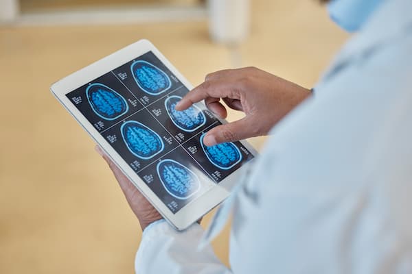
The analysis of MRI images is conducted by board-certified radiologists specializing in neuroradiology. These experts have extensive training in interpreting brain MRI scans and can identify abnormalities in brain structures, blood vessels, and soft tissues. They look for signs of neurological disorders, brain damage, tumors, or other conditions that may require further investigation or treatment. Their expertise ensures that you receive accurate diagnoses based on high-quality images from your MRI scan.
How to Access and Understand Your Results
Once your MRI images have been analyzed, the radiologist will prepare a detailed report outlining their findings. This report is typically sent to your referring physician, who will discuss the results with you.
You can request a copy of the report for your records. It’s essential to review this document carefully and ask questions if anything is unclear. Understanding the terminology used in the report can be challenging, so don’t hesitate to seek clarification from your healthcare provider.
Your MRI results may include terms like “lesion,” “mass,” or “abnormal signal,” which can be daunting. Your doctor will explain what these terms mean in the context of your health and what the next steps may be. They will also help you understand any implications for treatment or further testing that may be necessary based on the findings.
In summary, interpreting your MRI results involves collaboration between you, your radiologist, and your physician. This teamwork ensures that you have a clear understanding of your brain health and the best path forward for your care.
Why Southern Imaging Services is Your Best Choice
When it comes to obtaining an MRI of the brain, selecting the right imaging center is crucial. At Southern Imaging Services, we prioritize your health and comfort. Our facility is equipped with state-of-the-art MRI machines designed to provide high-quality images while ensuring a smooth experience for every patient.
Our board-certified radiologists specialize in neuroradiology, ensuring accurate diagnoses for conditions affecting the brain and spinal cord. They utilize advanced techniques in magnetic resonance imaging to capture detailed pictures of brain structures, blood vessels, and surrounding tissues. This expertise allows us to deliver precise assessments that are vital for effective treatment planning.
We understand that undergoing an MRI can be a source of anxiety for many patients. That’s why our patient-centric approach focuses on comfort and care. Our friendly staff is committed to making your visit as stress-free as possible.
We offer flexible scheduling, including same-day and weekend appointments, to accommodate your busy lifestyle. Our facilities are designed to provide a relaxing environment, with bilingual staff available to assist you in English or Spanish.
Choosing Southern Imaging Services means choosing a dedicated team that values your well-being. From the moment you walk through our doors to the moment you receive your results, we strive to provide the highest level of service. Trust us to handle your MRI scans with the care and professionalism you deserve.
Frequently Asked Questions
When considering an MRI for brain imaging, you may have several questions. Here are some common concerns we address:
Is the MRI procedure painful?
No, a brain MRI is a non-invasive imaging test. Most patients experience no discomfort during the scan. You will lie still inside the MRI machine, which may feel confined, but it is not painful.
How long does a brain MRI take?
Typically, a brain MRI scan lasts between 30 to 60 minutes. The exact duration can vary based on the specific imaging requirements and whether contrast material is used.
What if I am claustrophobic?
If you have concerns about claustrophobia, we offer open MRI options. These machines are designed to reduce feelings of confinement while still providing high-quality images.
Can I have an MRI if I have metal implants?
Certain metal objects, like some implanted medical devices, may interfere with the MRI process. It is crucial to inform your technologist about any implants, such as pacemakers or cochlear implants, before the exam. They will assess your situation and determine the best course of action.
MRI Safety and Considerations with Medical Devices
Safety is a top priority at Southern Imaging Services. Before your brain MRI, our MRI technologists will conduct a thorough screening process. This involves discussing your medical history and any devices you may have.
What devices are safe during an MRI?
Many devices, like removable dental work and some orthopedic implants, are generally safe during an MRI. However, items such as metal zippers, hearing aids, and electronic devices must be removed before the scan.
Are there any risks associated with MRI scans?
MRI scans do not use ionizing radiation, making them a safer alternative to CT scans. However, some patients may experience allergic reactions to contrast dye. It’s essential to discuss any allergies with your medical team before your exam.
What about pregnancy?
While MRIs are generally considered safe during pregnancy, we recommend consulting with your healthcare provider to weigh the benefits and risks before proceeding with the scan.
If you have any other questions or concerns about your upcoming MRI for brain imaging, don’t hesitate to reach out to our team for assistance. Your comfort and safety are our top priorities.
Contact Southern Imaging Services
Schedule Your MRI for Brain Scans Today
Southern Imaging Services is here to assist you if you need a brain MRI. Our team is dedicated to providing high-quality imaging services tailored to your needs. To schedule your MRI for brain imaging, simply reach out to us. You can call our office directly or use our online booking system for convenience. Our friendly staff will guide you through the process and answer any questions you may have.
Reach Out to Our Bilingual Support Team
At Southern Imaging Services, we understand the importance of clear communication. That’s why we offer support in both English and Spanish. Whether you have concerns about your MRI exam, questions about the procedure, or need assistance with scheduling, our bilingual team is ready to help. Don’t hesitate to contact us today. Your health and comfort are our top priorities.